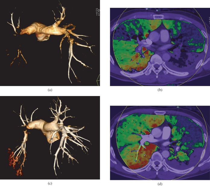
Figure 6.
Comparison between CT angiography and perfusion blood volume (PBV) images before and after thrombolytic therapy in a 39-year-old pulmonary embolism patient. (a, c) Volume reducing technique images show the disappearance of the right pulmonary artery emboli and significant reduction in the size of the left pulmonary artery emboli. (b, d) PBV images show improvement on the perfusion of the corresponding areas.

