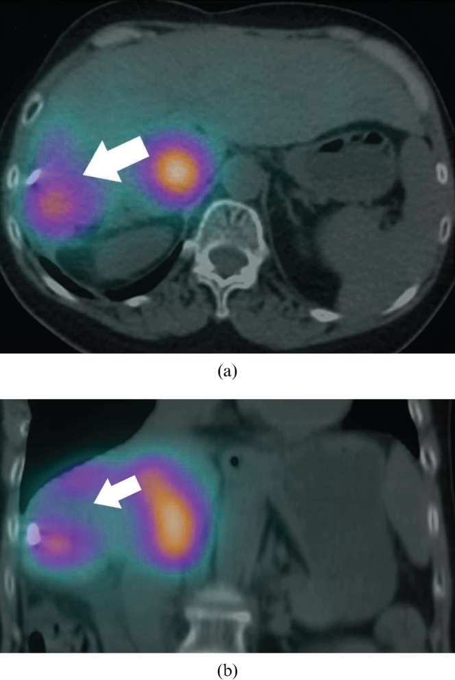
Figure 3.

Bremsstrahlung single-photon emission CT (SPECT)/CT acquired on the same day depicts bremsstrahlung activity as diffuse foci, inferior to the spatial resolution of yttrium-90 positron emission tomography/CT. Periablation cavity bremsstrahlung activity (arrow) cannot be clearly delineated. Bremsstrahlung activity in the caudate lobe is seen on SPECT/CT as a conglomerate focus with diffuse margins—(a) transaxial; (b) coronal.
