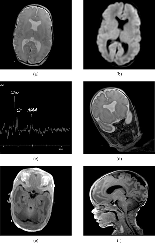
Figure 4.
MR images from a female born at 31 weeks′ gestational age by emergency Caesarean section for antepartum haemorrhage. (a) Axial fast spin-echo (FSE) T2 weighted image with the same parameters as Figure 3b. (b) Anatomically matched diffusion weighted image with 1 mm in plane resolution, 5 mm slice thickness, number of excitations = 1, repetition time/echo time = 10000/75.7 ms, b = 700 s mm–2. These do not show any evidence of hypoxic ischaemic injury. (c) Single voxel proton spectroscopy (shown with the same acquisition parameters as Figure 3e from the posterior hemispheric white matter) was normal. (d) Absence of the cavum septum pellucidum was confirmed, but in addition there was “squaring” of the frontal horns shown on coronal fast-recovery FSE T2 weighted images (shown with the same acquisition parameters as Figure 3b). This is characteristic of septo-optic dysplasia and hypoplasia of the optic nerves (especially the left side). (e) Chiasm was confirmed on axial reconstructions from an axially acquired volume data set (shown with the same acquisition parameters shown for Figure 3d). Opthalmology review confirmed bilateral optic disc hypoplasia, more pronounced on the left than the right. (f) Arrested migration of the posterior pituitary gland was confirmed on sagittal reconstructions of the volume data. This is a recognised associated finding in septo-optic dysplasia.

