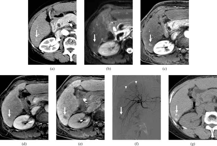
Figure 1.
A 68-year-old female with three (1.1, 2.3 and 2.5 cm in diameter) hepatocellular carcinomas (HCCs). (a) Contrast-enhanced CT scan obtained on arterial phase shows a faint enhancing nodule of 1.1 cm diameter with poor conspicuity in segment VI (arrow). The nodule showed no washout pattern on equilibrium phase (not shown). All observers interpreted the nodule as arterioportal shunt. (b) T2 weighted MR image shows a hyperintense nodule in segment VI (arrow). (c) Gadoxetic acid-enhanced arterial phase MR image shows a hypervascular nodule in segment VI (arrow). (d) Gadoxetic acid-enhanced 3-min late phase MR image shows the nodule with a washout pattern in segment VI (arrow). (e) Gadoxetic acid-enhanced hepatobiliary phase MR image shows a hypointense nodule in segment VI (arrow). All observers interpreted the nodule as HCC. (f) Right hepatic angiography shows a hypervascular tumour staining in segment VI (arrow). Other hypervascular tumour staining is also observed (arrowhead). (g) Unenhanced CT scan obtained after transcatheter arterial chemoembolisation shows iodised-oil accumulation at the corresponding HCC (arrow).

