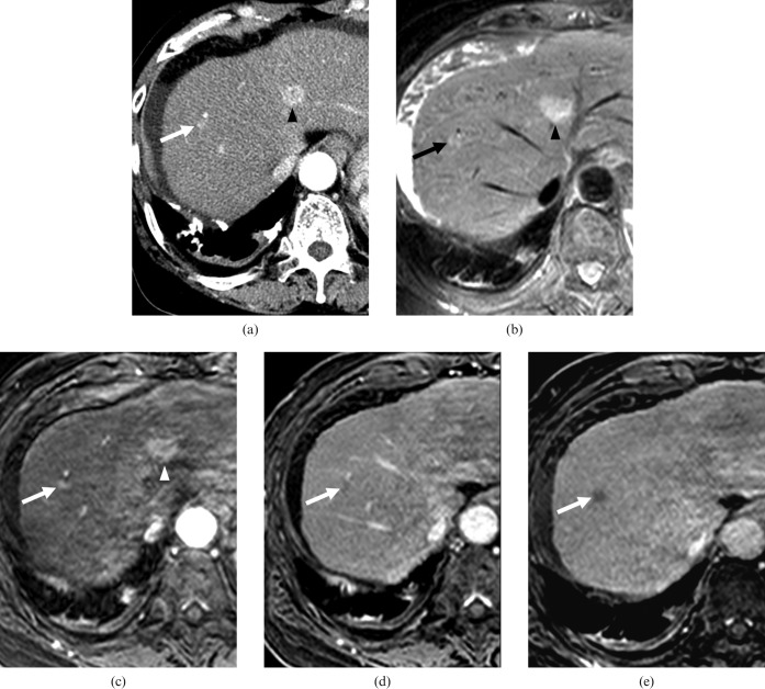
Figure 3.
A 64-year-old female with a dysplastic nodule in segment VIII and a hepatocellular carcinoma (HCC) in segment IV. The lesions were confirmed by a total hepatectomy with liver transplantation. (a) Contrast-enhanced CT scan obtained on arterial phase shows a hypervascular nodule of 0.9 cm diameter (arrow) in segment VIII and a hypervascular nodule of 1.8 cm diameter in segment IV (arrowhead). On contrast-enhanced CT scan obtained on equilibrium phase at the same level as (a), the nodule in segment VIII showed no washout pattern, as opposed to the nodule in segment IV, which did show a washout pattern (not shown). All observers interpreted the nodule in segment VIII as an arterioportal shunt, and the nodule in segment IV as HCC. (b) On T2 weighted MR image, two nodules in segments VIII (arrow) and IV (arrowhead) show hyperintensity. (c) Gadoxetic acid-enhanced arterial phase MR image shows two hypervascular nodules in segments VIII (arrow) and IV (arrowhead). (d) Gadoxetic acid-enhanced 3-min late phase MR image shows two nodules with a washout pattern in segments VIII (arrow) and IV (not shown). (e) Gadoxetic acid-enhanced hepatobiliary phase MR image shows two hypointense nodules in segments VIII (arrow) and IV (not shown). All observers interpreted two nodules as HCCs.

