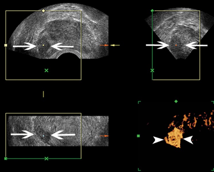
Figure 6.
An 80-year-old male suspected of having prostate cancer based on a borderline elevation in prostate-specific antigen (9.1 ng ml−1) level. Only two biopsy cores were positive in the right peripheral zone. Gleason score 4+4=8. Three-dimensional (3D) greyscale transrectal ultrasound shows an ill-defined hypoechoic nodule in the right peripheral zone on the transverse, sagittal and coronal planes (arrow). 3D power Doppler sonography shows distinct increased Doppler flow within the nodule (arrowhead).

