
Figure 2.
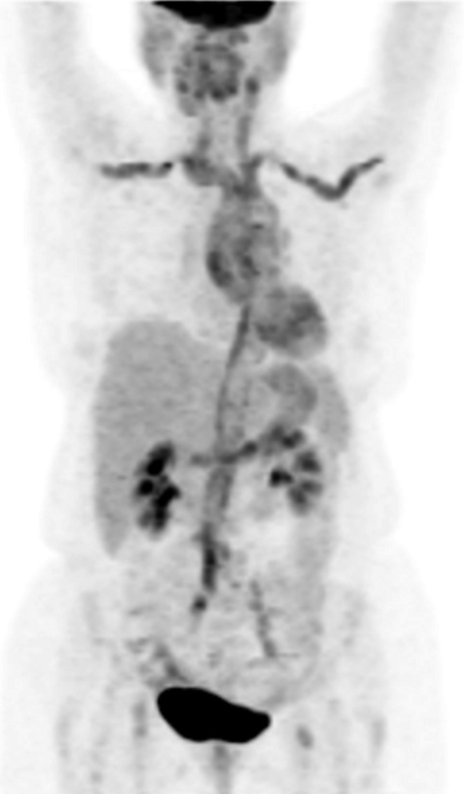
18F-Fludeoxyglucose (18F-FDG) positron emission tomography/CT (MIP image) in a 73-year-old female with giant cell arteritis at initial diagnosis. She presented with fever, malaise and increased inflammatory marker levels [erythrocyte sedimentation rate (ESR) 108, C-reactive protein (CRP) 96 mg dl–1]. There is intense 18F-FDG activity along the wall of the thoracic aorta and its tributaries, the subclavian and carotid arteries, the abdominal aorta and the iliac arteries. The degree of uptake was graded as 3, >liver.