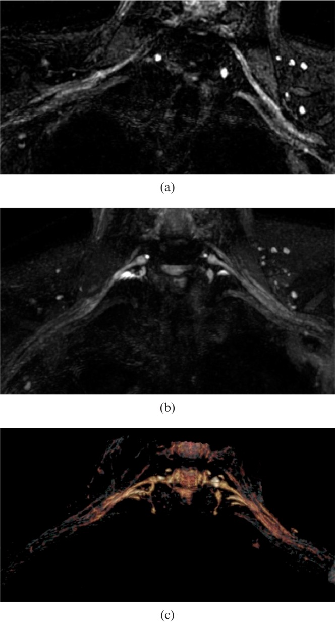
Figure 5.

Demonstration of brachial plexus fibres on a normal volunteer. Images are acquired with three-dimensional fast spin echo-cube with fat saturation. (a) The normal brachial plexus depicted on native images. (b) Fibres have been reconstructed using a multiplanar reconstruction algorithm. (c) Fibres are visualised in false colours using a volume rendering algorithm. Note that visualisation of roots is possible but not accurate.
