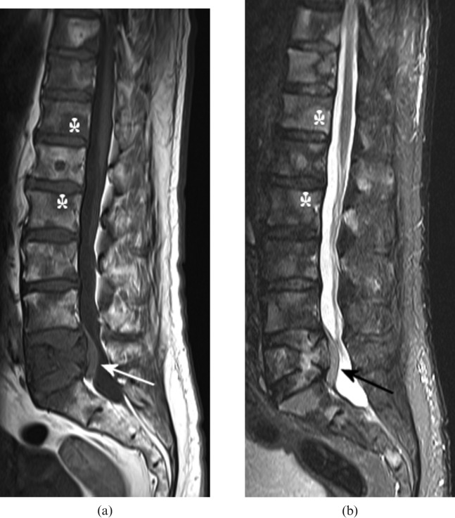
Figure 1.
(a) T1 weighted and (b) short tau inversion-recovery (STIR) MRI sequences of the lumbar spine in a 74-year-old male who presented with back pain. There are multiple vertebral lesions, which are of low signal on T1 weighting and high signal on STIR, consistent with tumour (asterisks indicate examples). L5 vertebral body is partially collapsed with a posterior soft tissue mass, which narrows the spinal canal and indents the thecal sac (arrows). Biopsy of an enlarged neck node showed non-Hodgkin lymphoma; the patient responded well to chemotherapy.

