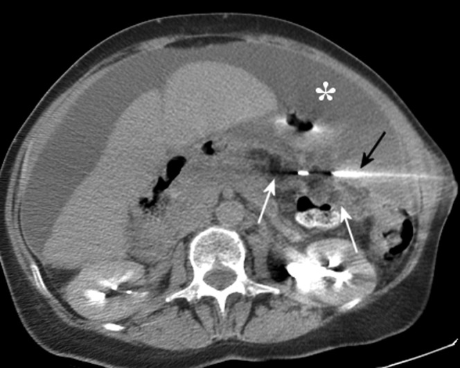
Figure 4.

CT-guided omental biopsy in a 63-year-old female who presented with abdominal distension and ascites (asterisk). The tip of the core biopsy needle (black arrow) is seen within the omentum of the left upper quadrant (white arrows), which shows soft tissue stranding consistent with tumour infiltration. In this case CT guidance was preferred to ultrasound guidance, owing to the deep position of the abnormality and the presence of overlying small bowel. Histology showed serous carcinoma with immunohistochemistry highly suggestive of an ovarian origin.
