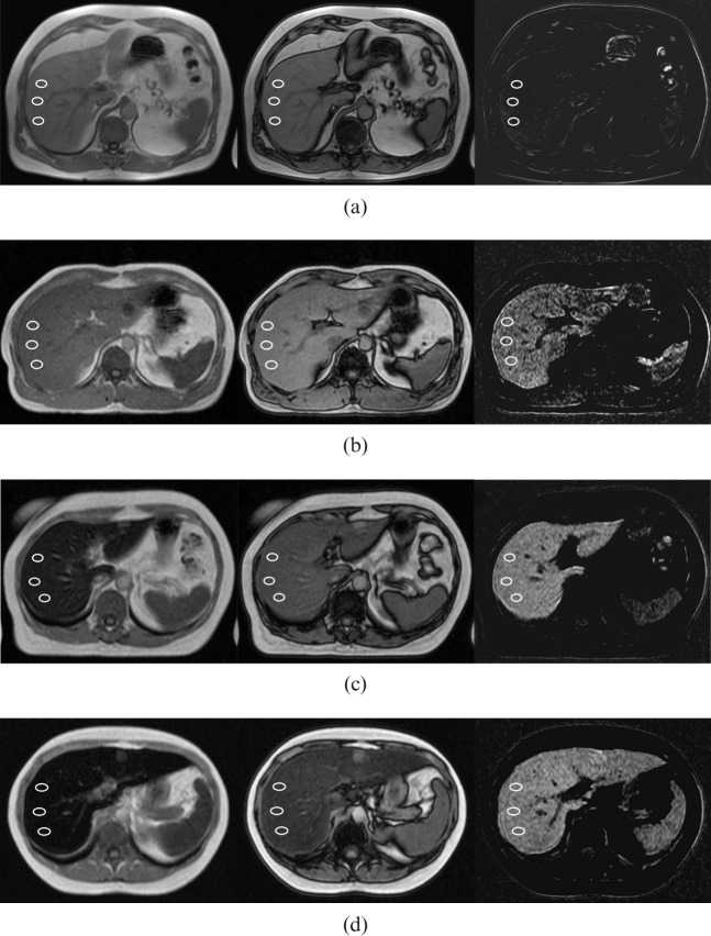
Figure 1.
T1 weighted axial in-phase and out-of-phase images demonstrating the criteria of visual grading and measurements of relative signal intensity (rSI) difference method in four patients as examples of different grades of iron overload. Three regions of interests were obtained for the rSI analysis. Left, in-phase image; middle, out-of-phase image; right, subtracted image calculated as the signal intensity of the in-phase image minus the signal intensity of the out-of-phase image. (a) Normal iron concentration (Grade 0) with MRI-based hepatic iron concentration (M-HIC) of 33 μmol g–1. (b) Minor iron overload (Grade 1) with M-HIC of 115 μmol g–1. (c) Moderate iron overload (Grade 2) with M-HIC of 193 μmol g–1. (d) Severe iron overload (Grade 3) with M-HIC of 271 μmol g–1.

