Figure 1.
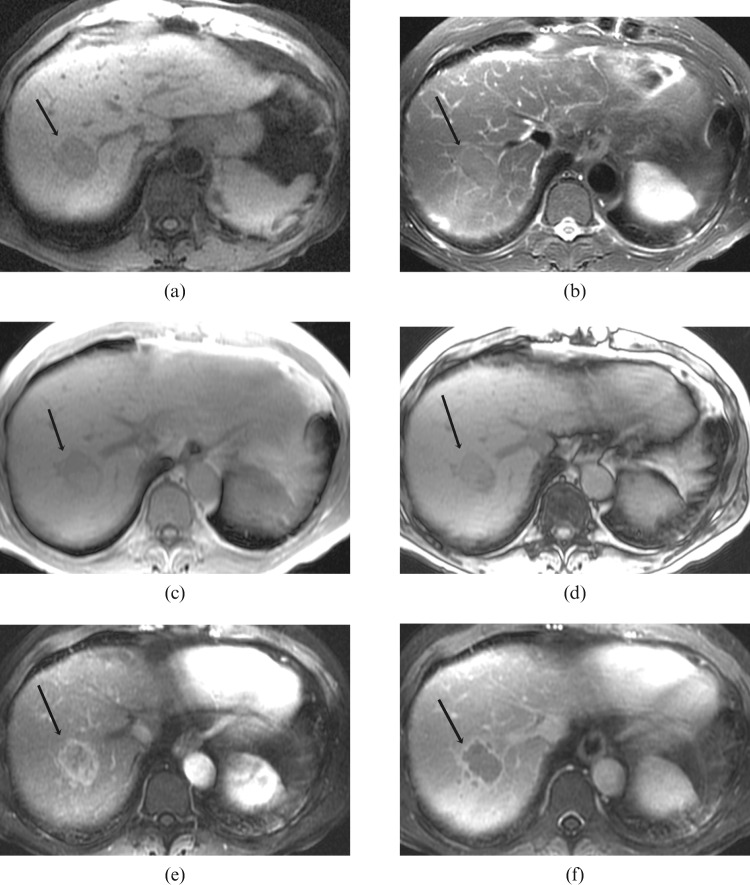
Histologically proven hepatocellular carcinoma in segment 7 of the liver in a 72-year-old male with relatively normal background liver texture: (a) axial T1 weighted image shows the lesion to be homogeneously hypointense to the rest of the liver with a well-defined margin (arrow); (b) axial T2 weighted image shows the lesion to be mildly hyperintense to the rest of the liver with a well-defined hyperintense rim (arrow); (c,d) axial T1 in- and out-of-phase images show signal dropout on the out-of-phase images in part of the lesion, in keeping with the presence of fat (arrow); (e) arterial T1 weighted image shows heterogeneous enhancement of the lesion with irregular rim enhancement (arrow); (f) portal venous T1 weighted image shows central washout with remaining irregular rim enhancement (arrow).

