Figure 2.

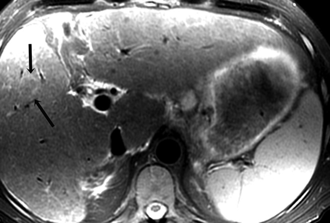
Histologically proven hepatocellular carcinoma in segment 5 of the liver in a 54-year-old male; axial T2 weighted image shows the lesion to be atypically hypointense to the rest of the liver with a well-defined hyperintense rim (arrows).

Histologically proven hepatocellular carcinoma in segment 5 of the liver in a 54-year-old male; axial T2 weighted image shows the lesion to be atypically hypointense to the rest of the liver with a well-defined hyperintense rim (arrows).