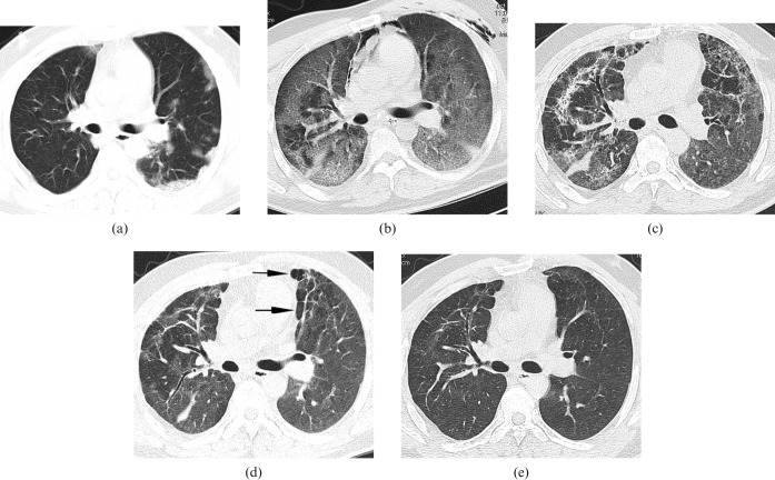
Figure 4.
Transverse thin-section CT scans in 34-year-old male with laboratory-confirmed swine-origin influenza A (H1N1). (a) Scan obtained on day 4 of illness shows a peripheral distribution of patchy ground-glass opacities (GGOs). With progression of symptoms, he was admitted to the intensive care unit on the sixth day of illness for mechanical ventilation. (b) Scan obtained on day 10 of illness shows diffuse bilateral GGOs; areas of consolidation predominately in subpleural and dependent lung regions. (c) Scan obtained on day 31 of illness shows irregular linear opacities that developed in the areas of GGOs. (d) Scan obtained on day 64 of illness shows that reticulation superimposed on GGOs persisted, although reduced in extent. Mosaic hypoattenuation (arrows) suggestive of air trapping in the ventral and subpleural lung regions can be seen. (e) Scan obtained on day 195 of illness shows resolution of fibrosis and air trapping.

