
Figure 4.
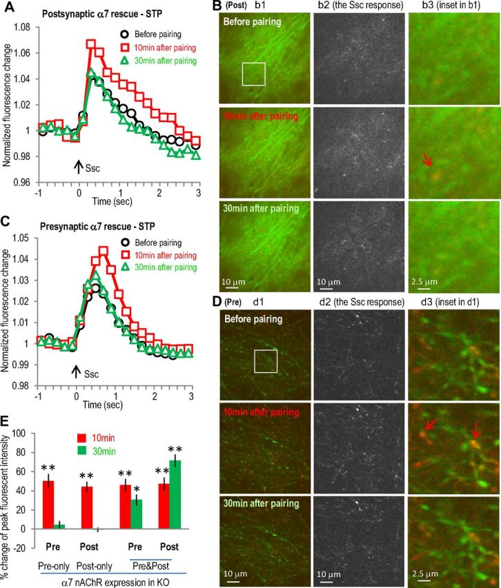
Expression of α7 nAChR in postsynaptic or presynaptic sites in α7 nAChR KO slice only induced transient activity enhancement in postsynaptic or presynaptic sites, respectively. A, α7 nAChR was coexpressed with GCaMP3 in hippocampal CA1 area of α7 nAChR knock-out mice (where the α7 nAChR-dependent LTP was abolished) as postsynaptic rescue of α7 nAChR. Normalized postsynaptic GCaMP3 responses show transient enhancement after the SO pathway pairing protocol. B, Images of postsynaptic calcium imaging from different time points before or after pairing protocol, with left panels showing the original images, middle panel showing the active calcium responses to SC stimulation, and right panels showing some potential dendritic spines. The potentiation was present in most of the dendrites responsive to SC stimulation (middle panels). C, α7 nAChR was coexpressed with GCaMP3 in hippocampal CA3 area as presynaptic rescue. Normalized presynaptic GCaMP3 responses show transient enhancement of presynaptic calcium activities after the SO pathway pairing protocol. D, Images of presynaptic calcium imaging from different time points before or after pairing protocol, with individual panels similar as described in B. E, Bar graph showing the requirement of both presynaptic and postsynaptic presence of α7 nAChRs to induce LTP. Red signals (in B and D) show the SC stimulation-induced calcium increases (also shown separately in the middle panels) over the basal signals before stimulation (green). Arrows indicate some synaptic sites with significant transient calcium increase. *p < 0.01, **p < 0.001, as compared with before pairing, Student's t test, n = 5 in each group. Ssc, Stimulation of the SC.