
Figure 1.
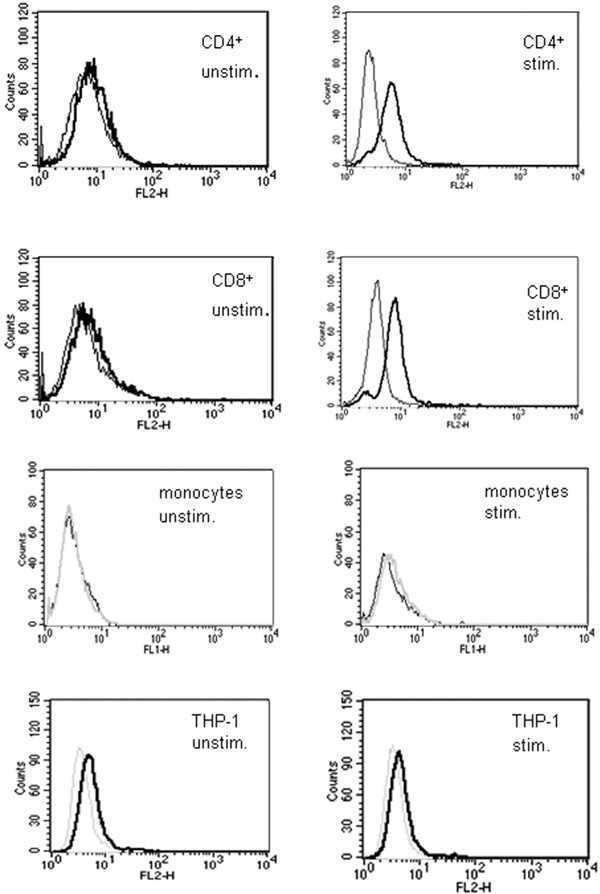
Surface expression of LIGHT on CD4+ and CD8+. Expression of LIGHT on the surface of CD4+, CD8+, CD14+ and THP-1 cells was measured by FACS. Cells were stimulated with PMA and ionomycin for 24 hours and stained with anti-LIGHT monoclonal antibody. LIGHT was not detectable on unstimulated cells, but activation of the cells resulted in appearance of LIGHT on the cell surface of CD4+ and CD8+ T cells, but not on monocytes and THP-1 cells. Thin curves correspond to the negative control and thick black curves correspond to specific staining for LIGHT. These data correspond to one representative experiment of three performed (Figure. 1).