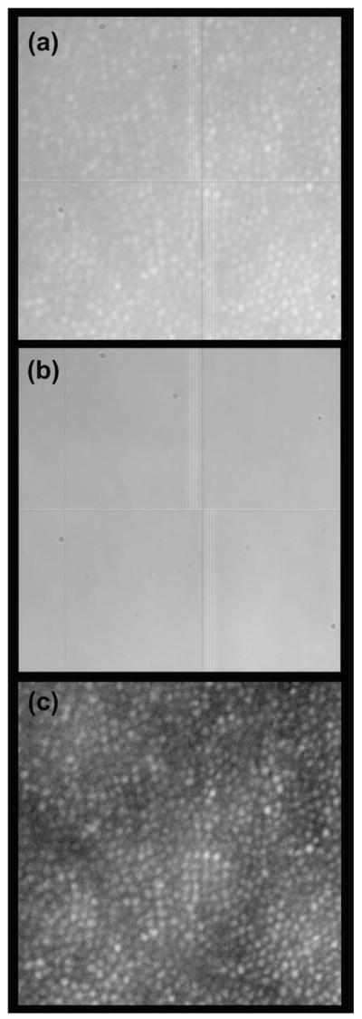
Fig. 2.

Processing retinal images from the Medical College of Wisconsin Adaptive Optics Ophthalmoscope. (a) Raw image from the CCD camera (0.5° × 0.5°). (b) Noise image comprised of dust, beam profile, and CCD circuit. (c) Processed retinal image (noise removed, single frame).
