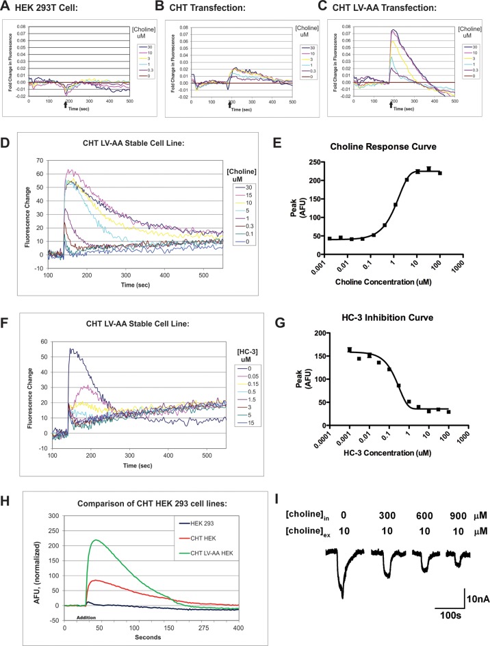
Figure 5.
Development of the CHT HTS assay using membrane-potential sensitive fluorescent dyes (Molecular Devices). Membrane potential sensitive dyes are incubated with HEK293 cells that are monitored in real time for fluorescent signal changes. (A) HEK293 cells do not generate a fluorescent signal following the application of choline (black arrow) and therefore do not demonstrate a choline-induced depolarization. (B–C) Transient transfection of WT CHT and CHT LV-AA into HEK293T cells demonstrated a specific choline-induced depolarization signal. (D–E) The LV-AA CHT stable cell line displayed an improved signal-to-noise ratio and a linear choline concentration–response from 0.1 to 30 μM. The signal measured using CHT LV-AA cells was significant enough to consider for the creation of a HTS assay to uncover allosteric potentiators of CHT. (F–G) The specific CHT antagonist HC-3 inhibits the choline-induced membrane depolarization of this cell line. (H) Summary comparison of the relative signal response of HEK293 cell lines expressing WT CHT and CHT LV-AA to choline-induced membrane depolarization. (I) CHT LV-AA expressing oocyte membrane current response to choline is reduced under conditions of very high internal choline concentrations.30.

