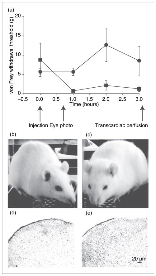
Figure 1.
Behavioral and immunohistochemical measures of the NTG rat model. (a) The threshold of von Frey hair for hindpaw withdrawal is displayed (mean ± SD) for NTG-treated (solid squares; n=5) and control rats (solid circles; n=5) after baseline acclimation. Indicated are the times of NTG or saline injection, eye photography, and transcardiac perfusion. (b) Saline and (c) NTG-treated rat representative of the consistent eye-squinting behavior. The vertical height:width ratio of the eyelids in three saline-treated rats was greater than those of three NTG-treated animals, p<0.0001, two-tailed t-test. (d) cFos expression in 40 μm thick sections of the lower brain stem showing the trigeminal nucleus caudalis (TNC) area, Sp5C41, after saline injection. (e) cFos expression in the TNC after NTG. Negative controls without primary cFos antibody had minimal cellular stain (data not shown).

