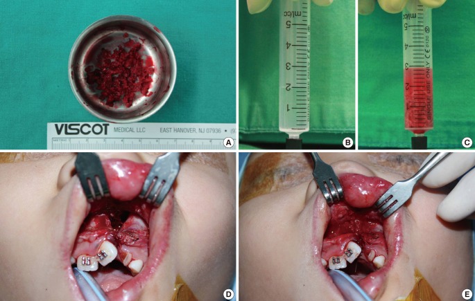
Fig. 3.
Intraoperative photograph
(A) The harvested cancellous bone chips approximately 2×2×2 mm in size. (B, C) The harvested bone was filled in syringe with 2 mL normal saline to measure the volume. (D) The alveolar bone defect prior to the cancellous bone graft. (E) The defect filled with cancellous bone chips. The cancellous bone was grafted in accordance with the maxillary contuor to avoid overcorrection.

