Figure 3.
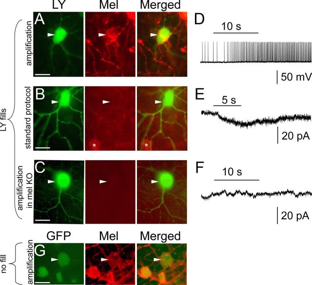
Immunohistochemical evidence for the presence of melanopsin protein in M4 cells. A–F, Tyramide-amplified, anti-melanopsin (Mel) immunofluorescence (red) is apparent in an M4 cell initially identified by its large, EGFP+ cell body (Opn4Cre/+;Z/EG+/− mouse) and further confirmed as an M4 cell based on its Lucifer yellow (LY)-filled dendritic arbor (green; arbor not shown) and intrinsic photosensitivity under synaptic blockade (A, D; current clamp; horizontal bar denotes the 10 s, 480 nm light stimulus). Melanopsin immunostaining is not apparent in an M4 cell from a mouse of the same genotype if tyramide signal amplification is omitted (B, E) or if tyramide amplification is used, but the melanopsin gene has been knocked out (C, F; Opn4Cre/Cre;Z/EG+/− mouse). Recordings in E and F were done in voltage clamp. G, A large M4 soma that was not filled or recorded expresses EGFP (green; left; Opn4Cre/+;Z/EG+/−), and melanopsin immunoreactivity using tyramide-amplification (red; middle). Collectively, these data indicate that melanopsin protein can be detected in M4 cells immunohistochemically provided the signal is amplified using tyramide (A). Arrowheads indicate location of M4 cells. Asterisks indicate neighboring melanopsin immunopositive ipRGCs, probably M2 cells. Scale bars, 20 μm.

