
Figure 3.
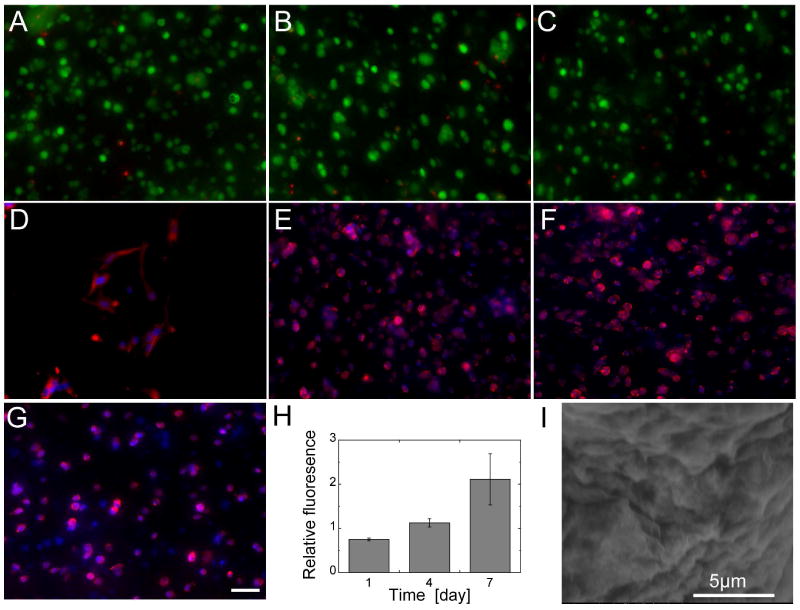
Live/Dead staining of the encapsulated mouse MSCs shows good viability at (A) day 1, (B) day 4, (C) and day 7. (D) Cells seeded on top of the hydrogel spread after one day, whereas cells encapsulated inside did not spread at (E) day 1, (F) day 4, (G) and day 7. (H) MTT assay shows an increased reduction over time, which indicates that cells are proliferating inside the hydrogel. (I) SEM image of hydrogel structure.