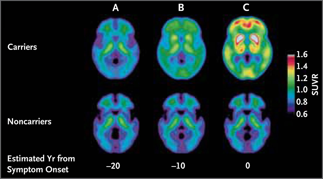
Figure 3. Aβ Deposition in Autosomal Dominant Alzheimer’s Disease Years before Expected Clinical Symptoms.
Panel A compares the fibrillar Aβ deposition, as measured by PET with the use of Pittsburgh compound B (PIB), of the average of autosomal dominant Alzheimer’s disease mutation carriers and noncarriers 20 years before the estimated time of onset of symptoms. There was significant Aβ deposition in the caudate and cortex in mutation carriers more than 10 years before expected symptom onset, as compared with noncarriers (Panel B). Panel C shows additional Aβ deposition throughout the cortex and neostriatum at the estimated time of symptom onset. An increased SUVR indicates increased binding of PIB to fibrillar amyloid. The scale ranges from low SUVR values (bluer colors), indicating low amounts of amyloid, to high SUVR values (redder colors), indicating high amounts of amyloid.

