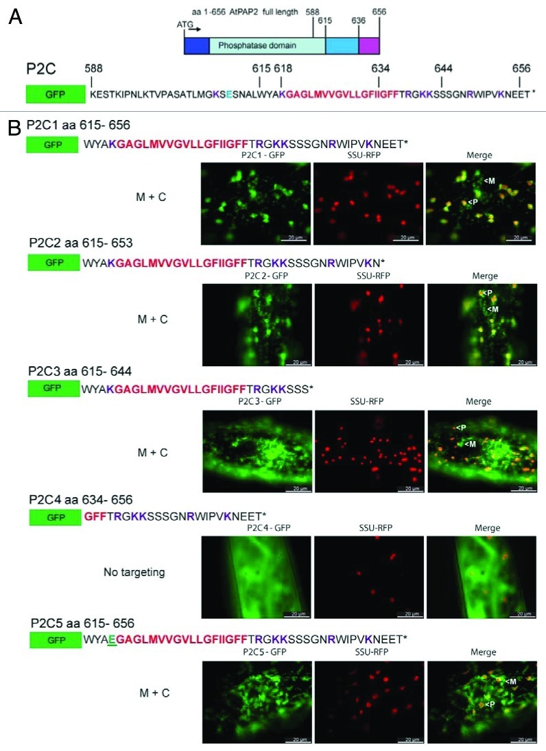
Figure 3. Targeting of GFP fused with various C-terminal extensions of AtPAP2. Hydrophobic motifs are in red; positively and negatively charged residues are in purple and blue, respectively; * denotes stop codon. (A) Full length AtPAP2 has been previously shown to be dual-targeted to the outer-membranes of plastids and mitochondria, by its C-terminal sequence.4 (B) A series of GFP constructs containing AtPAP2 with a number of modified C-terminal extensions were biolistically transformed into onion epidermal cells, alongside a plastidial RFP marker. Plastids and mitochondria have been identified in the merged micrograph by a (P) or (M), respectively. The K to E residue substitution in P2C5 has been shown in green.

An official website of the United States government
Here's how you know
Official websites use .gov
A
.gov website belongs to an official
government organization in the United States.
Secure .gov websites use HTTPS
A lock (
) or https:// means you've safely
connected to the .gov website. Share sensitive
information only on official, secure websites.
