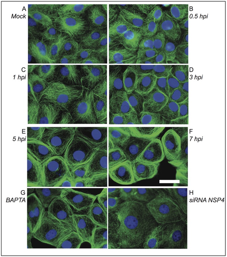
Figure 7. Changes in the tubulin cytoskeleton of MA104 cells infected with rotavirus.
Cells were either mock infected (A) or infected (B–H) with the reassortant rotavirus strain DxRRV at an m.o.i. of 10. At the indicated h.p.i. (B–F) cells were fixed and processed for immunofluorescence. Tubulin was visualized using a mAb anti-α tubulin as primary antibody and an anti-mouse FITC-conjugated secondary antibody (green). Nuclei were stained with DAPI (blue). Note that tubulin disorganization is apparent after 3 h.p.i. In addition, infected cells were treated with 25 µM BAPTA 2 h before fixation (G), or transfected with siRNANSP4 before infection (H) and fixed and processed for immunofluorescence at 7 h.p.i. Note that tubulin disorganization is partially or totally prevented in cell treated with BAPTA or transfected with siRNANSP4, respectively. Scale bar, 5 µm.

