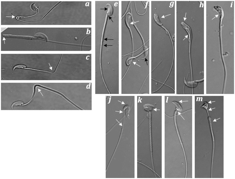
Figure 5. Morphological abnormalities of spermatozoa as seen in rescue lines expressing low levels of PPP1CC2.
(a, b, c, d) Mature epididymal spermatozoa with aberrant bending between mid-piece and principle piece. The angular variation of the bend ranges from 45° (a) to 90° (c, d) to 180° (b). (e–i) A series of frequently observed morphological defect involving jack-knifing of the head between the capitulum and the proximal mid-piece. (j–m) Other less common deformities, including shortened mitochondrial sheaths (j, m) and malformed heads (k, l, m). All micrographs were photographed under DIC optics with an Olympus 70 light microscope. Abnormal regions of the spermatozoa are denoted by white arrows and its corresponding normal regions are denoted by black arrows. These observations are representative of multiple samples from cauda epididymal preparations of different rescue animals.

