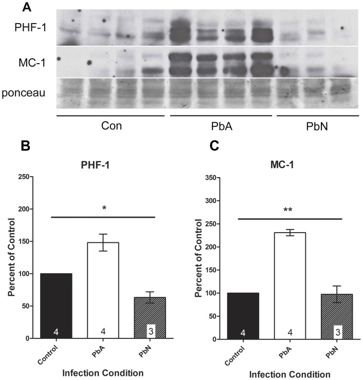
Figure 3. Abnormal tau expression in ECM.
Aberrantly phosphorylated tau was evident in PbA infected mice when compared to control and PbN infected mice. (A) Brain lysates were probed with antibodies to PHF-1 which stains for phosphorylated tau at Ser396/404, and MC-1 which recognized misfolded tau protein. (B) PbA infected mice demonstrated a 48% higher in PHF-1 immunoreactivity compared to controls and a 134% more compared to PbN. Significant group effects on the means was demonstrated by one-way ANOVA (F(2, 8) = 14.85; p<0.01) with significant mean differences between PbA mice and both control and PbN mice using post-hoc Tukey's multiple comparison test. Tukey's test did not demonstrate a significant effect of PbN infection on the mean PHF-1 expression when compared to control mice. (C) PbA-infected mice displayed a 131% more MC-1 expression compared to controls and 137% higher expression compared to PbN-infected mice. One way ANOVA demonstrated significant group effects in the expression of MC-1 (F(2, 8) = 51.29; p<0.001) with post-hoc Tukey's test demonstrating significant effect of PbA infection on MC-1 expression when compared to either control or PbN mice, but no effect of PbN infection when compared to controls. Densitometry measurements are illustrated as a percentage of corresponding measurements in uninfected controls. Values plotted as mean ± SEM. *p<0.05, **p<0.001. Con = control, PbA = P. berghei ANKA infected mice, PbN = P. berghei NK65 infected mice. Ponceau was used as loading control.

