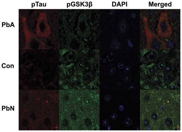
Figure 4. Immunofluorescence staining for phospho-tau (Red) and phospho-GSK3β (S9) (Green) in the brainstem.
PbA-infected mice displayed an increase in tau-positive neurons when compared to control mice or mice infected with PbN and an overall decrease or absence of intra-nuclear staining of pGSK3β (S9) in those neurons.

