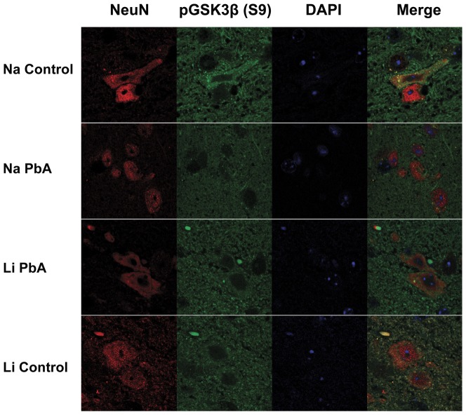
Figure 7. Immunofluorescence staining with NeuN (Red) and phospho-GSK3β (S9) (Green) in the brainstem.
PbA-infected mice displayed different patterns in the distribution of phospho-GSK3β within neuronal cells (NeuN-positive), with a lack of phospho-GSK3β in neuronal nuclei in contrast to LiCl treated ECM mice and uninfected control mice treated either with or without LiCl. Na Con = NaCl treated control, Na PbA = NaCl treated P. berghei ANKA infected mice, Li PbA = LiCl treated P. berghei ANKA infected mice, Li Con = LiCl treated control, NeuN = neuronal nuclear antibody.

