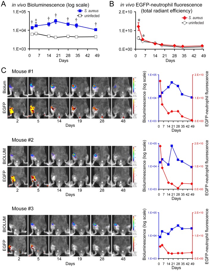
Figure 1. Two-dimensional in vivo bioluminescence and fluorescence imaging to monitor the bacterial burden and neutrophil infiltration during an orthopaedic implant infection.
S. aureus or no bacteria (uninfected) (n = 8 mice per group) were inoculated into the knee joints of LysEGFP mice in the presence of a titanium K-wire implant and mice were imaged using the IVIS Spectrum® imaging system (Caliper). (A) Mean bacterial burden as measured by in vivo bioluminescence (mean total flux [photons/s] ± sem) (logarithmic scale). (B) Mean neutrophil infiltration as measured by in vivo fluorescence (mean total radiant efficiency [photons/s]/[µW/cm2] ± sem). *p<0.05, †p<0.01, ‡p<0.001 S. aureus-infected mice versus uninfected mice (Student's t-test [two-tailed]). (C) (Left) Representative images of in vivo bioluminescence (upper panels) and in vivo fluorescence (bottom panels) overlaid on a grayscale photograph of the mice from the three S. aureus-infected mice that had different patterns of the bioluminescent signals. (Right) In vivo bioluminescent signals (photons/s/cm2/sr) (logarithmic scale) (blue) and in vivo fluorescent signals ([photons/s]/[µW/cm2]) (red) for each of these three mice.

