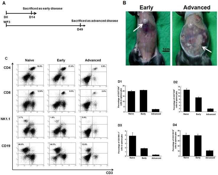
Figure 2. Alterations of systemic immune effector cells in splenocytes of mice challenged with PBS or WF-3 tumor cells.
(A) Diagrammatic representation of the collection of specimens in early and advanced diseases. (B) Representative figures of mice after WF-3 challenge at days 14 and 49. Note: Only small tumors (arrow) with little ascites were identified in mice of early disease. However, disseminated tumor implants (arrows) with bloody ascites within the whole peritoneal cavity were noted in mice with advanced disease. (C) Representative figures of flow cytometric analyses of various kinds of lymphocytes in splenocytes. (D) Percentages of various kinds of lymphocytes in splenocytes of naïve mice and in mice of early and advanced diseases. D1, CD4+ helper T lymphocytes; D2, CD8+ cytotoxic T lymphocytes; D3, NK1.1+ natural killer cells; and D4, CD19+ B lymphocytes. Note: All of the percentages of systemic immune effector cells significantly decreased as the tumor progressed from early to advanced stage.

