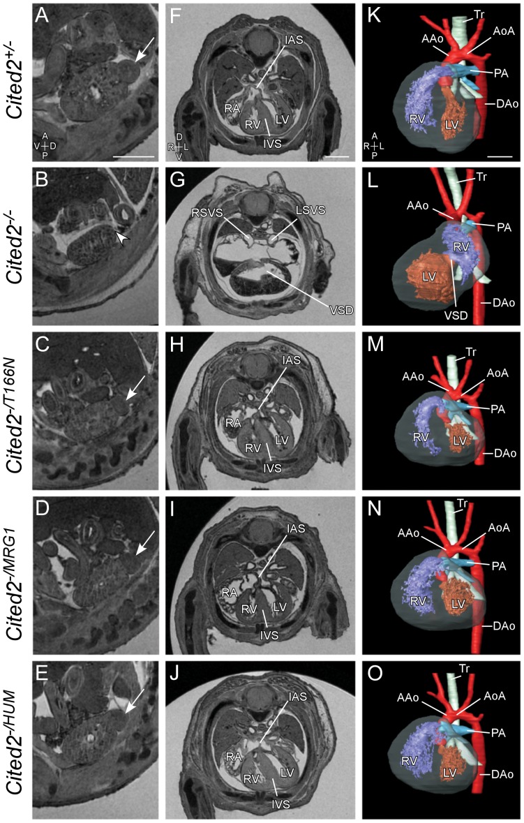
Figure 7. Phenotypic analysis of mouse embryos expressing CITED2 variants.
MRI analysis of embryos 15.5 days post coitum (dpc). Genotypes are indicated as shown. Sagittal sections through the left kidney (A–E) are shown to indicate the left adrenal gland where present (arrows), and absent (arrowhead). Transverse sections through the thorax (F–J) and 3D reconstructions (K–O) are shown to demonstrate cardiac anatomy. Loss of Cited2 leads to adrenal agenesis (B, arrowhead), right atrial isomerism, ventricular septal defect (VSD) and common atrium (G), and abnormal ventricular topology (L). Embryos expressing only the T166N variant, the MRG1 isoform, or full length human CITED2 have normal adrenal glands and hearts. RA, Right Atria; RV, Right Ventricle; LV, Left Ventricle; IVS, Interventricular Septum; IAS, Intra-atrial Septum; AAo, Aorta; AoA, Aortic Arch; Tr, Trachea; DAo, Dorsal Aorta; LSVS and RSVS, Left and Right Systemic Venous Sinus. Axis: A, Anterior; P, Posterior; V, Ventral; D, Dorsal; L, Left; R, Right. Scale bars: 0.5 mm.

