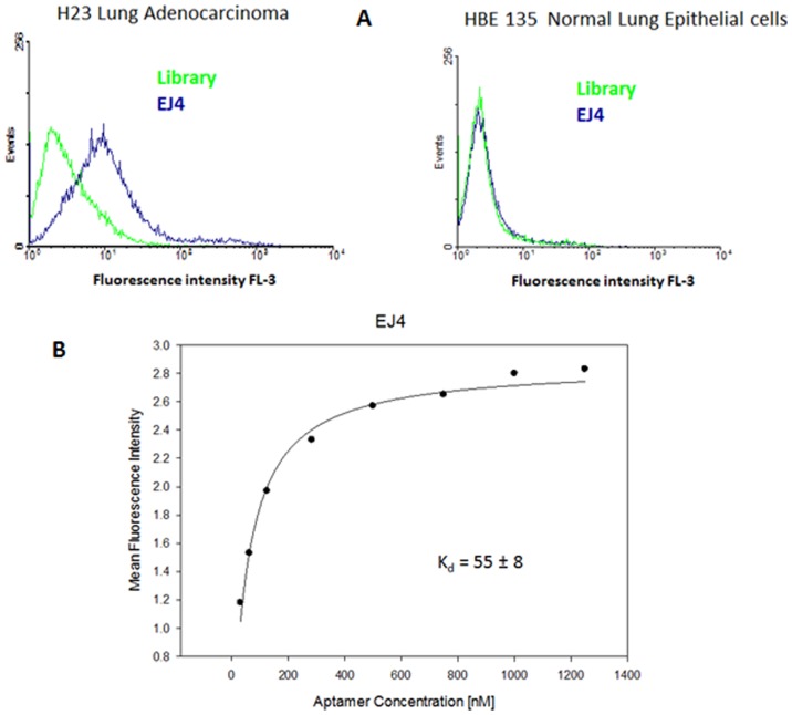
Figure 2. Characterization of selected aptamers.
Flow cytometry assay for the binding of the selected aptamer EJ4 with H23 (target cell line) and HBE135 E6/E7 (negative cell line). The green curve represents the background binding of a random sequence (library). Aim Figure 2: The aim of this figure is to show that the candidate aptamers are indeed aptamers because they do bind with the positive cell line (H23), but do not bind with the negative cell line (HBE135E6/E7).

