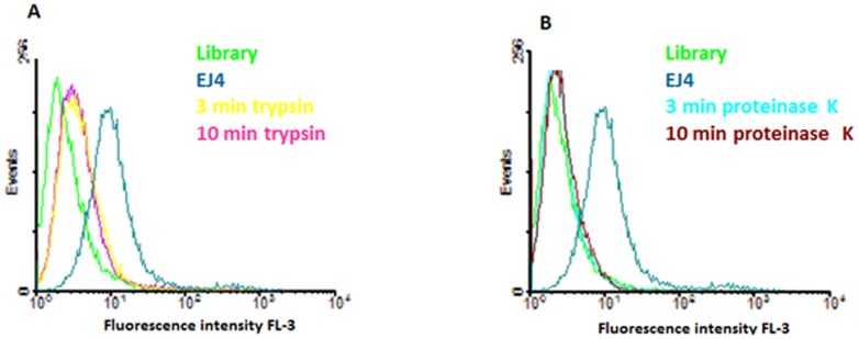
Figure 5. Binding Assays after proteinase treatment.
Flow cytometry assay for selected aptamers after treatment with proteases; untreated cells were used as positive control. (A) Cells treated with trypsin for 3 and 10 min prior binding with selected aptamer EJ4. (B) Cells treated with proteinase K for 3 and 10 min prior binding with aptamer EJ4. Aim figure 5: The aim of this figure is to prove that the targets of the selected aptamers are proteins present in the cell surface of the cancer cells (H23).

