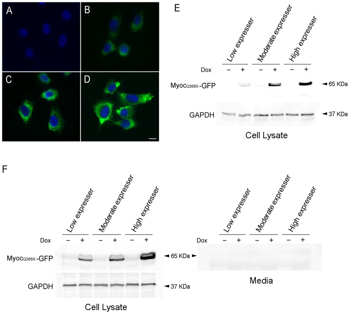
Figure 3. Tet-on inducible RGC5 cell lines that express mutant Q368X myocilin-GFP upon Dox induction.
Clones with different expression levels of Q368X myocilin-GFP (MYOCQ368X-GFP) fusion protein are presented. Without Dox induction, the green fluorescence in the cells was minimal (A). After Dox treatment, low (B), moderate (C) and high (D) levels of MYOCQ368X-GFP were seen in respectively, low, moderate, and high expresser clones. Cytoplasmic aggregates were visible in moderate and high expressers. Scale bar, 10 µm. E. Western blot analyses using anti-GFP and anti-GAPDH polyclonal antibodies. F. Western blot analyses of cell lysate (left panel) and medium (right panel) samples using anti-myocilin monoclonal antibody. The blots confirmed that the level of MYOCQ368X-GFP relative to that of GAPDH in total cell lysates was low, moderate, and high from the various expresser clones. No MYOCQ368X-GFP protein band was observed in medium samples. −, Non-induced control; +, Induced cells.

