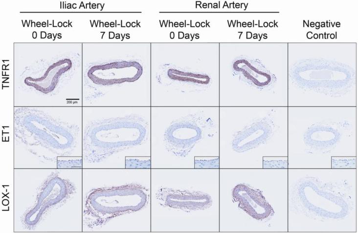
Figure 5.
Representative images of immunohistochemical staining (shown here in brown) for TNFR1, ET1, and LOX-1 in cross-sections of the iliac and renal arteries of wheel lock 0 days and 7 days rats. Pictures were captured at 20x magnification, whereas insets for ET1 were taken at 40x (scale bar = 50 microns). As illustrated, immunoreactivity of TNFR1 was abundant and it appeared homogenous across the arterial wall. In contrast, immunoreactivity of ET1was usually minimal and primarily localized in the endothelial cell layer. Immunoreactivity of LOX-1 generally appeared to be greater in the adventitia and endothelium, compared to the media. As summarized in text of the results section, mean data revealed no significant differences for any of the markers between groups.

