Figure 2.

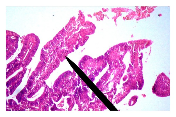
Photomicrograph (400x, H&E stain) of bronchoscopic biopsy showing papillary configuration lined by malignant cells along with prominent fibrovascular core (pointer).

Photomicrograph (400x, H&E stain) of bronchoscopic biopsy showing papillary configuration lined by malignant cells along with prominent fibrovascular core (pointer).