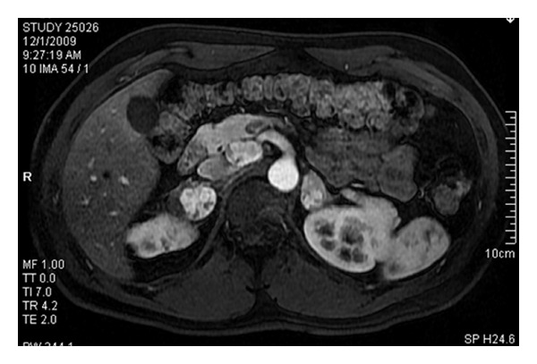
Figure 3.

Arterial phase T1-weighted image in the axial plane following intravenous contrast depicts hypervascular adrenal masses on both sides and an approximately 3 cm hypervascular mass in the uncinate process of the pancreas consistent with a neuroendocrine tumor.
