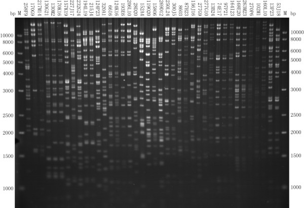
Figure 2.
A representative image of DNA fingerprints of the positive BAC clones for determination of overlapping relationship. The positive BAC clones identified in the previous steps were digested with Hind III, followed by separation on a 1% agarose gel in 1× TAE buffer. The gel was stained with Ethidium Bromide (EB) for photograph with a UVP Labworks system. M: Marker of DNA size standard (1 kb plus DNA ladder from Invitrogen, San Diego, CA, USA) with the base pair (bp) sizes indicated on both sides.

