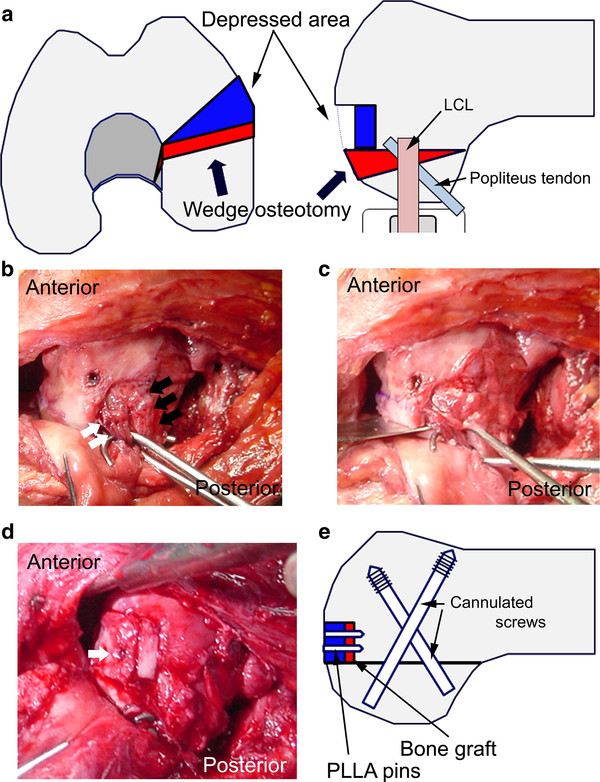
Figure 3.
a) Plan for 5-mm closed wedge osteotomy and elevation of the depressed portion. b) Photograph of the lateral aspect of the left knee. The 2 skin hooks retracted the popliteus tendon (white arrow), and lateral collateral ligament (LCL) (black arrow). c) The skin hook retracted these structures to protect them from the bone saw during osteotomy. d) The depressed portion was elevated by using a bone graft and fixed using poly-l-lactic acid (PLLA) pins (white arrow). e) Schema after wedge osteotomy and internal fixation.

