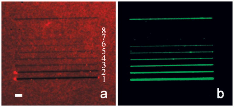
Figure 3.

Epifluorescence images of (a) a nanoshaved BSA monolayer and (b) SLB lines. The top line, which is ~200 nm in width, was used as a reference marker. The widths of shaved lines in (a) from 1 to 8 are ~600, ~300, ~142, ~103, ~78, ~55, ~36, and ~15 nm, respectively, as measured by AFM. The length of each line is 40 μm. The scale bar is 3 μm. Note, the vacant lines in (a) become increasingly difficult to observe by epifluorescence microscopy as the line width narrows. Green fluorescence emanating from these regions, however, should be trivial to observe even for the thinnest lines under the conditions of this experiment, if a bilayer is indeed present.
