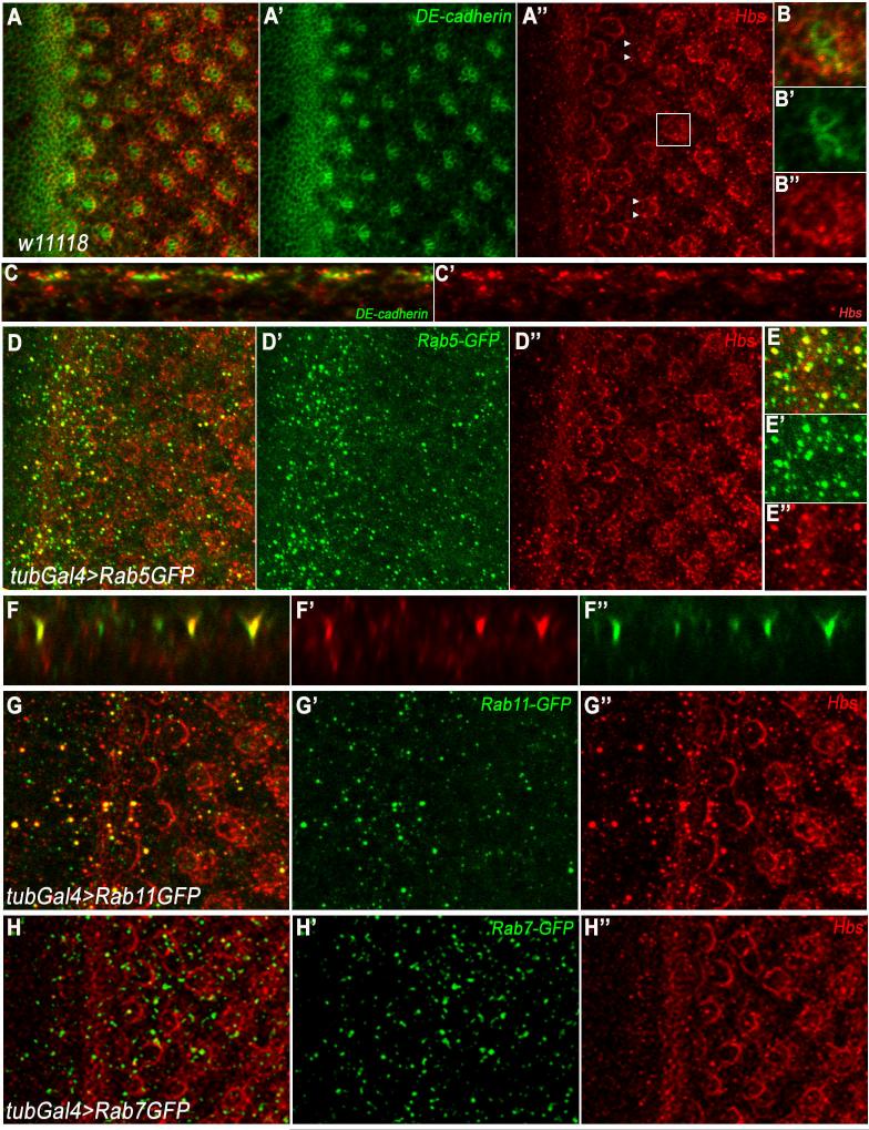
Figure 2. Hbs is localized at membranes/junctions and in intracellular puncta.
(A-B) Confocal projections depicting localization of Hbs (red) and DE-cadherin (green, marking cellular outlines at junctional level and highlighting developing photoreceptor clusters) in third instar eye discs. Hbs is upregulated in the furrow (MF, white arrow in A and A”) and R-cell preclusters, as they emerge from MF. Hbs is enriched at membranes surrounding ommatidial preclusters (A”, magnified in B-B”), reflecting expression in all R-cells.
(C-C’) x/z-section of third instar eye disc in A. Note that Hbs is not only enriched in junctional membranes but also present in intracellular puncta throughout R cells.
(D-E) x/y-section of third instar eye disc of tubGal4>Rab5GFP genotype, stained for Hbs (red) and Rab5GFP (green). Note Hbs positive puncta co-localize (magnified in E-E’) with Rab5GFP puncta, a marker for early endosomes.
(F-F’) x/z-section of third instar eye disc in D, showing co-localization of Hbs and Rab5 positive puncta.
(G-G’) tubGal4>Rab11GFP eye discs showing co-localization of Hbs (red) with recycling endosomal marker Rab11 (green).
(H-H’) tubGal4>Rab7GFP eye discs showing expression of Hbs (red) and Rab7GFP (green). Hbs does not co-localize with late endosomal marker Rab7 (green).
See also Suppl. Figure S2.

