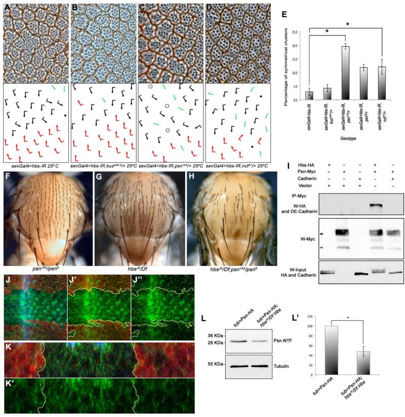
Figure 6. Hbs is required for Psn function and processing.
(A-D) Tangential adult eye sections of indicated genotypes, anterior is left and dorsal up (arrows and dots as in Figure 1). sevGal4>hbs-IR (A) is not affected by kuze29-4/+ heterozygosity (B), whereas dosage reduction of psn (sevGal4>hbs-IR, psn143/+) (C), and nct (sevGal4>hbs-IR, nctA7/+) (D) strongly enhances the hbs-IR phenotypes, suggesting a positive relationship between Psn and Hbs. (E) Quantification of R3-R3 symmetrical clusters in genotypes shown in B-D. Error bars represent standard deviations (**p<0.001).
(F-H) Adult nota of genotypes indicated, anterior is up. (F) heteroallelic hypomorphic psn143/psn9 combination with normal mechanosensory bristle arrangement; (G) hbsJ5/Df(2R)ED2423 mutant thorax displaying occasional “bald” patches as a result of missing bristles; and (H) hbsJ5/Df(2R)ED2423; psn143/psn9 double mutant thorax with many bristles missing, suggesting synergistic interaction and hbs and psn acting in the same molecular context.
(I) Hbs is co-immunoprecipated (co-IP) by Psn: immunoblot from S2 cell whole cell lysates expressing Hbs-HA either alone or in combination with Psn–Myc or DE-Cadherin (acting as negative control). Cell lysates were immunoprecipitated with anti-Myc (IP-Myc) and blots were probed with anti-HA (Hbs) and anti-DE-Cadherin antibodies, revealing specific Co-IP of Hbs with Psn while DE-Cadherin (a control protein similar to Hbs) does not bind to Psn-Myc (bottom panel: Hbs input). See also Suppl. Fig. S5I for additional controls.
(J-J”) Confocal projections of third instar larval eye discs stained for Psn-HA (green), DE Cadherin (blue, labeling apical surfaces of all cells), and lacZ (red, marking wild-type tissue: hbs mutant tissue marked by absence of red; outlined by yellow lines in J’-J”). hbs mutant tissue shows marked accumulation of PsnHA. (K-K”) Transverse (x/z)-section of eye disc shown in (J); note accumulation of Psn in the hbs mutant tissue (marked by absence of lacZ).
(L-L’) Western blot of third instar larval eye-brain complexes showing Psn-NTF fragment in wild-type and hbs mutant larvae. In hbs mutant larvae there is a marked reduction of Psn-NTF fragment (quantified in L’) as compared to control flies (γ-Tubulin, lower panel, serves as loading control). Error bars represent standard deviations, with *p<0.001.
See also Suppl. Figure S5.

