
Figure 6.
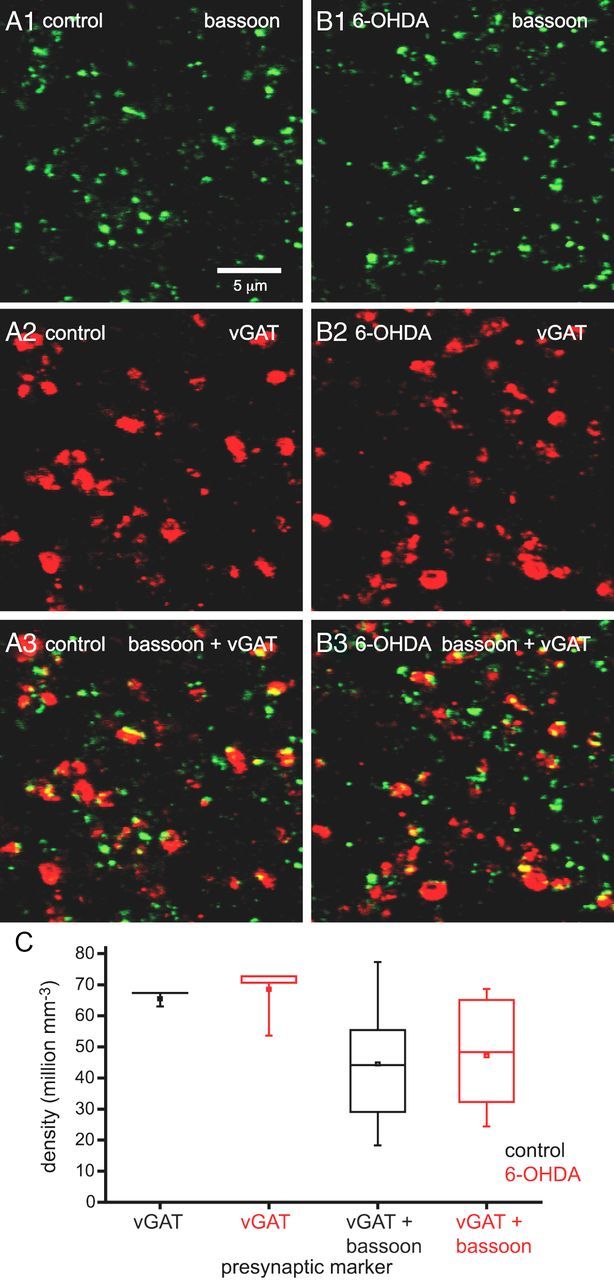
Chronic dopamine depletion did not alter the densities of presynaptic vGAT- and bassoon-immunoreactive structures. A1–B3, Representative confocal micrographs of bassoon (A1, B1: green), vGAT (A2, B2: red), and bassoon plus vGAT immunoreactivity (A3, B3: yellow) in the STN of a control (A) and 6-OHDA-depleted (B) animal. The densities of immunoreactive structures in the STN were not altered by chronic dopamine depletion compared with control in the representative micrographs (A, B) or across the sample population (C). The scale bar in A1 applies to each micrograph. *p < 0.05.