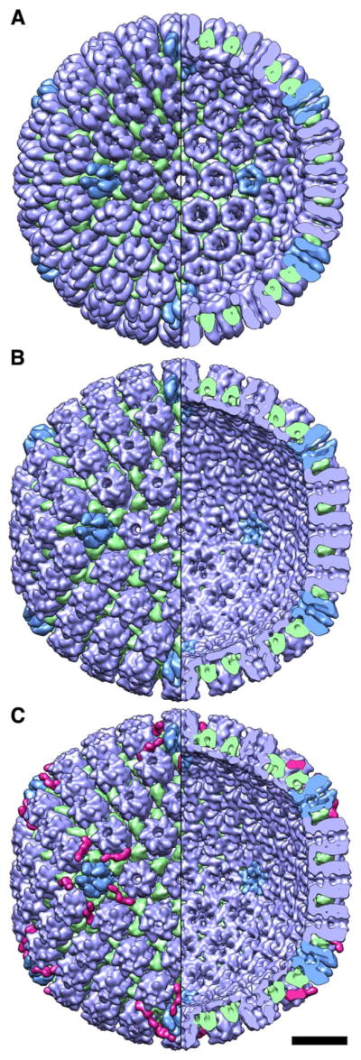
Figure 2. Cryo-EM reconstructions of HSV-1 capsids.

(A) the HSV-1 procapsid; (B) A-capsid; and (C) C-capsid, with DNA computationally removed. Left half of each reconstructon shows the outer surface and right half, the inner surface. Bar, 20 nm.

(A) the HSV-1 procapsid; (B) A-capsid; and (C) C-capsid, with DNA computationally removed. Left half of each reconstructon shows the outer surface and right half, the inner surface. Bar, 20 nm.