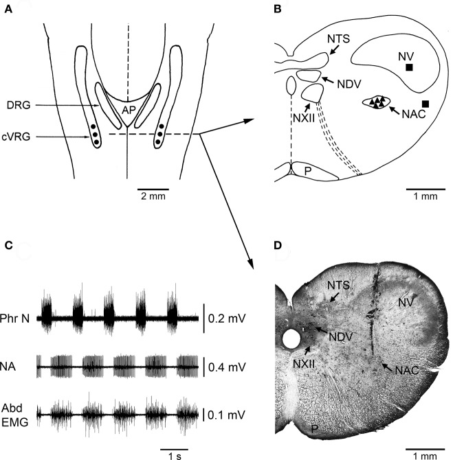
Figure 1.
Localization of injection sites and histological control. (A) A diagrammatic representation of a dorsal view of the medulla oblongata of the rabbit showing where bilateral microinjections of bicuculline or muscimol were performed into the cVRG (•). AP, area postrema; cVRG, caudal ventral respiratory group; DRG, dorsal respiratory group. (B) Diagram of a coronal section of the medulla oblongata at the level indicated in panel A (dashed line) showing the location of representative sites (▲) where the microinjections of 1 mM bicuculline were performed. The diagram also shows the location of some control injection sites (■) where bicuculline caused no appreciable changes in the pattern of breathing and cough responses. NAC, nucleus ambiguus caudalis; NDV, nucleus dorsalis nervi vagi; NTS, nucleus tractus solitarii; NV, nucleus tractus spinalis nervi trigemini; NXII, nucleus nervi hypoglossi; P, tractus pyramidalis. The atlas of Meessen and Olszewski (1949) was used for comparison. (C) An example of neuronal expiratory activity recorded in the cVRG region. Phr N, phrenic neurogram; NA, neuronal activity; Abd EMG, abdominal electromyographic activity. (D) Photomicrograph of a coronal section of the medulla oblongata at approximately the same level as in B showing the location of a track along which a 1 mM bicuculline microinjection was made into the cVRG. Intense multiunit expiratory activity was recorded at this injection site.

