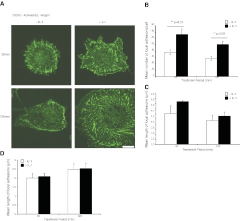
Figure 3.
A) HGFs plated on FN-coated glass coverslips (30 or 120 min) and treated with vehicle or with IL-1β (40 ng/ml) were immunostained for activated β1 integrin (12G10 antibody) and visualized by TIRF microscopy. Scale bar = 10 μm. B) Mean ± sd numbers of focal adhesions per cell that were immunostained with 12G10. Data are from measurements of >10 cells/group. C) Mean ± sd length of focal adhesions immunostained with 12G10 in the cell body (>5 μm from the cell membrane; >10 cells/group). D) Mean ± sd length of focal adhesions in the cell periphery (<5 μm from the cell membrane; >10 cells/group). Focal adhesions were quantified with MetaMorph.

