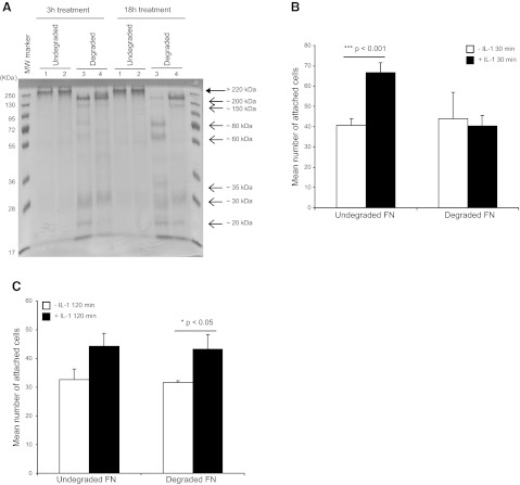
Figure 7.
A) FN was attached to plates and incubated with buffer (undegraded) or MMP3 at 1:10 or 1:100 enzyme-FN substrate ratios for 3 or 18 h (degraded). Native FN or FN-degradation fragments were separated by SDS-PAGE. Native FN (large arrow; >220 kDa) and degraded FN fragments (small arrows: ∼200-20 kDa) were stained with Coomassie blue. Lane 1, untreated FN; lane 2, native FN incubated with buffer alone; lane 3, FN digested with MMP3 at enzyme-substrate ratio of 1:10; lane 4, FN digested with MMP3 at enzyme-substrate ratio of 1:100. B, C) HGFs were plated on native FN or MMP3-digested FN (at 1:10 ratio) for 18 h and treated with vehicle or with IL-1β (20 ng/ml) for 30 min (B) or 120 min (C). FN was washed with PBS before cell adhesion assays. Adherent cells were counted by enumeration of DAPI-stained nuclei in 100-μm2 grids. Mean ± sd numbers of attached cells were calculated from values obtained from 4 wells.

