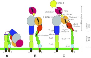
Figure 1.
Schema of LFA-1 conformation. A) LFA-1 in a resting condition. Legs are in a bent conformation. B) Leg-extended conformation with a closed headpiece; the cytoplasmic domains are separated. KIM127 epitope is shown as a black circle. C) Leg-extended conformation with an open headpiece. Arrows next to the α7 helices in the αI and βI domains indicate that both helices are pulled down. Both the αI and βI MIDAS are shown as black stars. MEM148 epitope is shown as a black circle.

