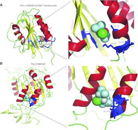
Figure 4.
Docking of isoflurane on the αI and βI domains using Glide. A) Docking of isoflurane to the αI domain in the closed conformation (PBD 1ZOO). B) Docking of isoflurane to the βI domain of β2 subunit in the closed conformation (PBD 3K6S). Residues photolabeled by azi-isoflurane are shown in blue. Right panels show blowup of the docking site. Asterisks indicate αI MIDAS. In isoflurane: red, oxygen; green, chloride; light blue, fluoride.

